
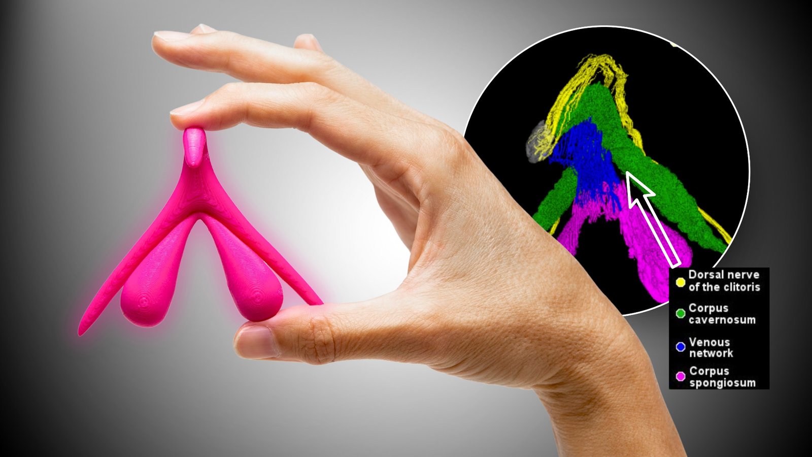
Researchers have produced a highly detailed 3D view of key sensory nerve pathways inside the clitoris.
Using HiP-CT, Ju Young Lee and colleagues followed the dorsal clitoral nerve as it branched through the glans. Five major nerve trunks appeared in both postmortem specimens.
The imaging also showed some branches extending toward the clitoral hood and mons pubis. Posterior labial nerves were seen supplying areas surrounding the clitoris and labia.
That level of detail is the real advance. Earlier studies had already traced clitoral nerves, but fine branches inside the glans are difficult to follow through conventional dissection.
Both specimens were postmenopausal. Autonomic nerves were outside the research scope, and the paper remains a preprint.
The researchers say the added anatomical detail may help inform surgery around the vulva. That is where the study becomes relevant to Filipino clinicians and patients too. Better anatomical information can matter wherever procedures take place near these nerve pathways, even though improved patient outcomes have not yet been demonstrated.
Calling it a map of “all the nerves” goes further than what the study found. Researchers produced a much finer view of key sensory pathways, but they did not map every clitoral nerve.
A two-specimen preprint can reveal anatomy. Is that enough to influence surgery, or should clinical practice wait for much stronger evidence?
Source • Lee et al., bioRxiv preprint, 20 March 2026